Absorbance, Transmittance, and Analytical Principles

Prepare Sample

Dissolve your sample in a suitable solvent  and put it in a special transparent container (cuvette). Also, prepare a reference cuvette (called blank) with pure solvent. 

Shine Light

The machine shines a beam of UV and visible light through both the sample and the reference.

Select Wavelength (Scanning Type)

A part of the machine (monochromator) acts like a filter, selecting one specific color (wavelength) of light at a time to pass through the sample. It then repeats this for all the wavelengths in the UV/Vis range.

Measure Light

A detector measures how much of the light passes through the sample and how much passes through the reference at each wavelength.

Calculate Absorbance

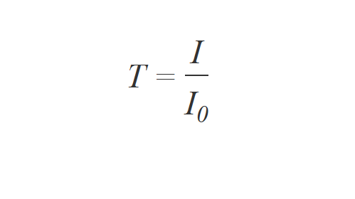
The machine compares the amount of light that passed through the sample to the amount that passed through the reference. This tells us how much light the sample absorbed at each wavelength.

Show Results

The machine then creates a graph (the UV/Vis spectrum) showing how much light the sample absorbed at each different wavelength. This helps identify and quantify the substances in the sample

Standard Macro Cuvettes

These are the most common type and are suitable for most samples. They typically have external dimensions of 12.5 mm x 12.5 mm and a height of 45 mm, with internal dimensions of 10 mm x 10 mm, resulting in a standard path length of 10 mm.

Long Path Cuvettes

These cuvettes have a path length greater than 10 mm and are used when a sample is too diluted, requiring a longer path length to increase the absorbance signal and improve sensitivity. They are also useful when a sample may vaporize or undergo a chemical change during measurement, as the longer path allows for more interaction with the light beam.

Short Path Cuvettes

Cuvettes with a path length of less than 10 mm are used when the absorbance of a sample is very high and dilution is either difficult or undesirable. The shorter path length helps keep the absorbance within the instrument's measurable (linear) range.

Micro Cuvettes

These cuvettes are specifically designed for analyzing very small sample volumes. For example, some micro cuvettes feature an optical path length of 10 millimeters and are made of fused quartz glass. They are suitable for measurements in the ultraviolet and visible range, covering wavelengths between 200 nm – 2500 nm, and can handle sample volumes around 700 µL.

Flow Cells

They are suitable for measurements in the ultraviolet and visible range, covering wavelengths between 170 nm – 2700 nm, and require a small sample volume of 440 µL. These cells are reliable and reusable.

Calibration involves checking and adjusting the accuracy of the instrument in the following areas. These checks are conducted regularly, and adjustments are made if the readings fall outside acceptable limits, making it essential to keep records of these calibrations.


Wavelength

Ensuring that the selected color of light is correct by using standard reference materials.

Absorbance/Transmittance

Confirming that the instrument accurately measures the amount of light absorbed or transmitted through standard solutions.

Stray Light

Checking for any unwanted light that could lead to measurement errors.

Resolution

Verifying the instrument's ability to distinguish between closely spaced colors of light.

Baseline

Ensuring a stable and accurate zero reading.

Food and Beverage

Ensures consumer safety by assessing food quality and composition, focusing on attributes like color, flavor, and aroma. It also employs analytical techniques to identify contaminants and adulterants.

Pharmaceutical

Rigorous analysis is vital for verifying the purity, concentration, and identity of drugs. Monitoring the stability of medications is also essential to ensure efficacy over time under varying environmental conditions.

Cosmetics

Evaluates product safety and effectiveness by analyzing the photostability of UV filters, characterizing particles, and measuring color indices. It also detects adulteration and quantifies dyes and antioxidants to meet consumer expectations.

Petrochemical

Characterizes crude oil, calculates asphaltene fractions, formulates aromatic content indices, determines sulfur content, and calculates solubility factors.

Chemical

Determines chemical properties, assesses final product quality, studies polymer composition, qualifies water, determines purity and dyeing efficiency, analyzes photocatalytic degradation and pesticide residues.

Biotechnology

Determines concentration and purity of nucleic acids and proteins, monitors microbial cell cultures, studies protein denaturation and kinetics, and analyzes biological samples like blood plasma and serum.

UV/VIS Applications

UV/VIS Applications

Search for specific applications.

color measurement with UV Vis

Color Measurement

Understanding Color Measurement: Importance, Techniques, and Applications

UV/VIS water testing

UV/Vis Water Testing

Discover the Role of UV/Vis Spectrophotometry in Achieving Accurate Water Quality Analysis

Spectrophotometry Applications and Fundamentals Guide

Spectrophotometry Applications and Fundamentals Guide

Guide on Fundamental Knowledge as Well as Application Tips and Hints

Anthocyanogens in Beer - UV Vis Spectrophotometry

Anthocyanogens in Beer - UV Vis Spectrophotometry

Determination According to Harris and Ricketts

Vitamin B12 Analysis - UV Vis Spectroscopy

Vitamin B12 Analysis - UV Vis Spectroscopy

Peak Identification of Cyanocobalamin using a UV Vis Spectrophotometer

 Hydroquinone in Cosmetics

Hydroquinone in Cosmetics

Determination using UV/VIS Spectroscopy at 289 nm

APHA Color Number

APHA Color Determination - UV Vis Spectroscopy

APHA Color measurements of near-clear samples according to CIE

Carbonyl in Aldehyde & Ketone - UV Vis Spectroscopy

Carbonyl in Aldehyde & Ketone - UV Vis Spectroscopy

Spectroscopic Determination of Total Carbonyl Content

BCA Protein Assay - UV Vis Spectroscopy

BCA Protein Assay - UV Vis Spectroscopy

Colorimetric Quantitation of the Total Protein Concentration in Biological Samples

What are the different types of spectroscopy?

Type of Spectroscopy

Type of Radiation

Interactions

Wavelength

ϒ-ray spectroscopy

Y-rays

Atomic nuclei

< 0.1 nm

X-ray fluorescence spectroscopy

X-rays

Inner shell electrons

0.01 – 2.0 nm

Vacuum UV spectroscopy

Ultraviolet (UV)

Ionization

2.0 – 200 nm

UV/Vis spectroscopy

UV/Vis

Valance electrons

200 – 800 nm

Infrared & Raman spectroscopy

Infrared

Molecular vibrations

0.8 – 300 mm

Microwave spectroscopy 

Microwaves

Molecular rotations

1 mm – 30 cm

Electron spin resonance spectroscopy

Electron spin

Nuclear magnetic resonance spectroscopy

Radio waves

Nuclear spin

0.6 – 10 

The different spectroscopic techniques are mainly differentiated by the radiation they use, the interaction between the energy and the material, and the type of material and applications they are used for. The spectroscopic techniques commonly used for chemical analysis are atomic spectroscopy, ultraviolet and visible spectroscopy (UV Vis spectroscopy), infrared spectroscopy, Raman spectroscopy and nuclear magnetic resonance.


How do you read a UV/Vis spectrum?

To read a UV/Vis spectrum, you analyze the plot of absorbance (or sometimes transmittance) versus wavelength. Key points to focus on include:

  • Absorption Peaks: Identify the wavelengths where the absorbance is highest; these correspond to electronic transitions in the molecules.
  • Peak Wavelengths (λmax): The wavelength(s) at which maximum absorbance occurs, characteristic of specific molecular structures or functional groups.
  • Peak Intensity: The height of the absorption peaks indicates how strongly the substance absorbs at that wavelength, related to concentration and molar absorptivity.
  • Baseline: Check the baseline absorbance in regions without absorption to assess instrument or sample issues.
  • Shape of Spectrum: The shape and number of peaks can provide information on the types of electronic transitions and the environment of the molecules.

By interpreting these features, you can identify compounds, determine concentration, and study molecular properties.

What is the range of UV/Vis spectroscopy?

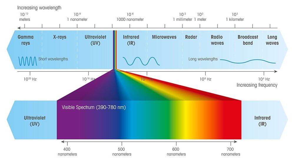
UV/Vis spectroscopy typically covers the wavelength range from 190 nm to 780 nm. 

More specifically: 

  • The ultraviolet (UV) region generally ranges from 190 nm to 390 nm.
  • The visible (Vis) region generally ranges from 390 nm to 780 nm.

METTLER TOLEDO’s UV/VIS Excellence spectrophotometers extend further into the near-infrared region, reaching 1100 nm.

Why is UV/Vis spectroscopy important and why is it used?

UV/Vis spectroscopy is important because it enables both qualitative and quantitative analysis of substances by measuring their absorption of ultraviolet and visible light.

This method helps determine the concentration of analytes, study chemical kinetics, assess purity, perform objective color measurements, and analyze molecular structures. Its applications cover a wide range of fields, including chemistry, biology, environmental science, and materials science, making it a versatile and essential tool for analysis.

What is the difference between fluorescence spectroscopy and UV/Vis spectroscopy?

UV/Vis spectroscopy measures the light absorbed by a sample to determine its concentration and identify compounds. This process involves the removal of light.

In contrast, fluorescence spectroscopy measures the light emitted by a sample after it absorbs light, usually at a longer wavelength. This technique focuses on the re-emission of light, providing much higher sensitivity for specific fluorescent molecules.

How do you prepare a sample for UV/Vis spectroscopy?

To prepare a sample for UV/Vis spectroscopy, you'll need to handle your cuvettes and solutions carefully:

  1. Prep the Cuvette: Securely place your cuvette in a rack; don't fill it while it's in the instrument.
  2. Add Solutions: First, pipette your blank solution into one clean cuvette. Then, pipette your sample into a separate clean cuvette. Always use plastic pipette tips to avoid scratching the cuvette.
  3. Fill Level: Fill the cuvette to a maximum of 4/5 full, avoiding underfilling or overfilling.
  4. Clean & Check: Wipe the cuvette to remove any droplets or fingerprints. Before measuring, ensure no air bubbles are inside and that your sample is homogeneously mixed.

How do you determine the concentration of an unknown solution using UV/Vis spectroscopy?

To determine the concentration of an unknown solution using UV/Vis spectroscopy, follow these steps:

  1. Prepare a calibration curve: Measure the absorbance of a series of standard solutions with known concentrations at the wavelength of maximum absorbance (λmax) for the analyte.
  2. Plot absorbance vs. concentration: Create a calibration curve by plotting the absorbance values against the known concentrations. According to Beer's Law, absorbance is directly proportional to concentration.
  3. Measure the unknown sample: Record the absorbance of the unknown solution at the same λmax.
  4. Determine the concentration: Use the calibration curve to find the concentration corresponding to the measured absorbance of the unknown sample.

This method relies on Beer-Lambert Law, which states that absorbance A=εbc, where ε is the molar absorptivity, b is the path length, and c is the concentration.

What are the different molecular interactions in the UV region?

Types of Transition in UV Region

The absorption of UV light results in electronic transitions from lower energy levels to higher energy levels. Absorption of ultraviolet radiation in organic molecules is restricted to certain functional groups (chromophores) that contain valence electrons of low excitation energy. The molecular transitions/interactions that take place due to UV absorption are:

  • π- π* (pi to pi star transition) – bonding to anti-bonding orbital
  • n - π* (n to pi star transition) – non-bonding to anti-bonding orbital

These transitions need an unsaturated group in the molecule to provide the π electrons.

σ (bonding) to σ* (anti-bonding) transitions require higher energy and therefore cannot be detected using UV Vis spectroscopy.

How do functional groups affect the spectra?

Consider a functional group containing atoms with one or more lone pairs of electrons that do not absorb ultraviolet/visible radiation. However, when this functional group is attached to a chromophore, it alters the intensity and wavelength of absorption. This phenomena is called an auxochrome or a color-enhancing group.

The presence of an auxochrome causes the position shift of a peak or signal to a longer wavelength, which is called a bathochromic or red shift. The functional groups contributing to bathochromic groups are substituents such as methyl, hydroxyl, alkoxy, halogen and amino groups.

The auxochrome that causes position shift of a peak or signal to shorter wavelength is called a hypsochromic or blue shift. Actually, the combination of chromophore and auxochrome behaves like a new chromophore having a different absorption maxima (λmax). For example, benzene shows λmax at 256 nm, whereas aniline shows λmax at 280 nm. Hence, the NH2 group acts as an auxochrome and causes the shift of λmax to a larger value.

What Is the difference between spectral bandwidth and resolution in UV/Vis spectroscopy?

InstrumenSpectral resolutionEquivalent SBW (nm)
UV5> 1.5< 2.0
UV5Bio> 1.5< 2.0
UV5Nano> 1.7< 1.5
UV7> 1.9≤ 1.0

The table shows the resolution of METTLER TOLEDO's UV/VIS Excellence spectrophotometers, which is measured using toluene in hexane, and the equivalent SBW.

The spectral bandwidth (SBW) of a spectrophotometer is related to the physical slit-width and optical dispersion of the monochromator system. Resolution is the ability of an instrument to separate light into finite, distinct wavelength regions and to distinguish each finite region. Spectral bandwidth is typically used for scanning instruments, whereas resolution is typically used for array instruments.

For most pharmacopeia quantitative purposes, a spectral bandwidth of less than 2 nm is sufficient and the acceptance criteria for the ratio is 1.3. Spectral resolution can be used for comparison with spectral bandwidth.

What are the different light sources used in a UV/Vis spectrophotometer?

Light Source

Wavelength Range

(nm)

RegionLifetime
Tungsten filament lamp350 – 2500Vis + IR3,000 hr
Deuterium arc lamp190 – 400UV1,000 hr
Hydrogen lamp190 – 400UV1,000 hr
Xenon flash lamp190 – 1100UV + Vis + NIR5,500 hr*

* Corresponds to 50 Hz flashes at constant operation

The best light source would be one that provides good intensity with low noise across all ultraviolet and visible wavelengths and offers stability over a long period. There is a range of light sources which are commonly employed as mentioned above.

How is diffraction grating better than a prism?

Diffraction gratings are generally better than prisms for splitting different wavelengths because:

Higher Spectral Resolution: Gratings can provide much higher spectral resolution due to their ability to produce multiple sharp diffraction orders, allowing finer separation of closely spaced wavelengths.

Linear Dispersion: The angular separation between wavelengths in a grating is more linearly related to wavelength, making it easier to analyze and calibrate spectra compared to the nonlinear dispersion of prisms.

No Material Dispersion Limitations: Prisms rely on material dispersion (variation of refractive index with wavelength), which can limit performance, especially in certain wavelength ranges. Gratings use interference effects, which are not limited by material properties.

Broad Wavelength Range: Gratings can work efficiently over a broader range of wavelengths, including ultraviolet and infrared, whereas prisms have absorption and dispersion limitations.

Overall, diffraction gratings provide more precise and versatile wavelength separation than prisms, which is why they are commonly used in spectrometers and optical instruments.

Which inorganic compounds can be measured by UV/Vis Spectroscopy?

Molecules can be analyzed using UV/Vis spectroscopy if they possess any functional group or conjugation, or if they produce a color complex. As inorganic compounds do not contain any functional group or conjugation, the common method for analyzing them is by reaction with a suitable compound. This produces a color complex whose absorbance can be photometrically measured in the visible region and correlated with its actual concentration. For example, iron is commonly analyzed by a reaction with 1, 10-phenthroline to produce a red color complex. The absorbance of the complex is measured at 570 nm to estimate iron concentration.

How do single beam and double beam spectrophotometers differ?

The main difference between a single beam and double beam spectrophotometer follows.

  • Single beam spectrophotometer: A single beam from the light source passes through the sample
  • Double beam spectrophotometer: The light beam from the light source is split into two parts: one part goes through the sample, and the other part passes through the reference

Beam splitting in a double beam spectrophotometer is achieved in two ways:

  1. statically, with partially transmitting mirrors or a similar device
  2. attenuating the beams using moving optical and mechanical device

How to analyze solid polymer film using UV/Vis?

The analysis of a solid sample is performed mainly by estimating its absorbance, transmittance and reflectance. Common parameters determined for solid polymers include % transmittance, cutoff wavelength, and yellowness index. The sample is mounted on a holder specifically designed for solid samples and readings are taken in the same manner as they are for liquid samples. A solid sample holder enables measuring of solid samples such as films or glass.

solid sample holder

Does temperature affect UV/Vis analysis?

Temperature affects absorbance values. Different solvents undergo different interactions at different temperatures. Solution parameters that change due to temperature changes are:

  • Rate of reaction. The rate changes when temperature is elevated. This can cause a change in the activity of the sample. Enzymatic/biomolecular reactions are very sensitive to temperature.
  • Solubility of a solute. Solubility is affected with variations in temperature. Poor solubility may result in imprecise absorption.
  • Expansion or contraction of the solvent. This may lead to a change in the concentration of the solution and affect the absorbance, as absorbance is linearly related to concentration.
  • Schlieren effect. This effect may occur with temperature changes, leading to a series of convective currents which may change the true absorbance.

Optical performance parameters such as photometric noise, wavelength accuracy/repeatability, photometric repeatability and stray light are not influenced by temperature within a range of 10 – 40 °C.

Whereas, optical parameters like photometric resolution (toluene/hexane ratio) and photometric accuracy wavelengths (K2Cr2O7 in HClO4) show a temperature dependency ranging from 0.014 to -0.034/unit within 10 – 40 °C.

What is stray light?

What is Stray Light?

Stray light is defined as light that reaches the detector which is not  from the instrument's light source and does not follow the optical path, causing a deviation at the corresponding wavelength. Therefore, the light intensity measured by the detector is higher than it actually should be. Conversely, this also means that the measured absorbance is lower than the true absorbance because it is reduced by the contribution of stray light. This effect is more prominent at higher absorbance values (high sample concentrations).

Download the whitepaper to learn more about the origin and accurate measurement of stray light:

Stray Light and Performance Verification

Why is the sample compartment in UV/Vis array spectrophotometers open?

The sample compartment in UV/Vis array spectrophotometers is open due to the fact that array instruments use reverse optics and the simultaneous detection of all wavelengths of the spectrum.

  • Reverse optics: The light is diffracted after it has gone through the sample. Due to this, only a small fraction of the external ambient light contributes to the signal in a given wavelength region.
  • Simultaneous detection: Using an array detector which provides 2048 light intensity signals at the same time, full spectrum is recorded within one second. Because the measurement is very fast, the effect of ambient light is significantly reduced.

I want to…
Need assistance?
Our team is here to achieve your goals! Speak with our experts.